
New technique maps protein interactions within intact human tissues
UChicago PME researchers developed a method to simultaneously profile genes, proteins, and protein complexes across tissues, opening new windows into immunity and disease
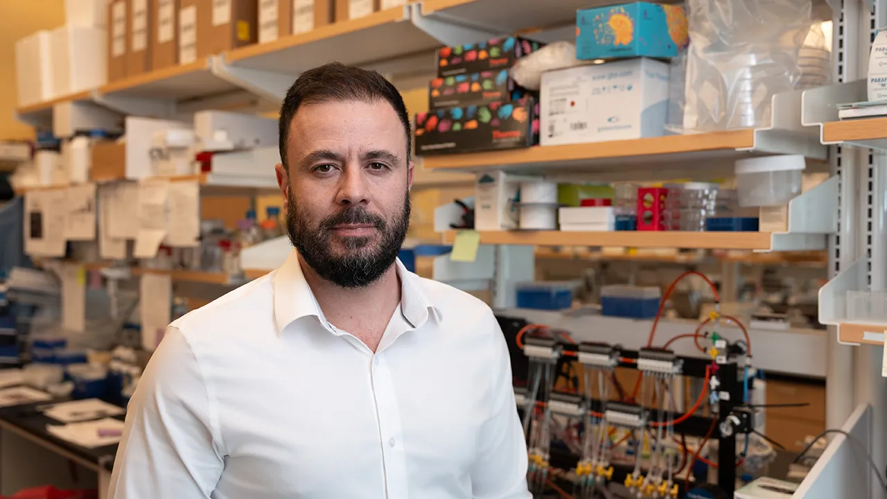
A new study from the lab of UChicago PME Prof. Savaş Tay describes a technique to simultaneously measure proteins, protein interactions, and gene expression in intact sections of tissue. (Photo by Jason Smith)
The proteins that carry out nearly every function inside our bodies rarely act alone. As they send messages between cells, recognize pathogens, and build the machinery that keeps cells alive and dividing, proteins work in teams. But most methods to capture the state of a cell have relied on quantifying gene activity or overall levels of proteins, failing to capture how any proteins present are actually interacting.
Now, researchers at the University of Chicago Pritzker School of Molecular Engineering (UChicago PME) have developed a technique that can shed light on exactly which proteins are interacting with which other proteins—and where.
Called Spatial Proximity-Sequencing, or Sprox-seq, it simultaneously measures proteins, protein interactions, and gene expression in intact sections of tissue. In a new study published in the journal Cell, the team applied Sprox-seq to human tonsil tissue, profiling 32 proteins, 528 possible protein interactions, and thousands of genes that guide immune cell behavior.
“To truly understand how cells function together as tissues, we need to know not just which proteins are present, but how they are actually communicating with each other,” said UChicago PME Prof. Savaş Tay, corresponding author of the new work. “Sprox-seq gives us a way to capture those molecular conversations alongside gene activity, and to see exactly where in the tissue they are happening.”
Sprox-seq gives us a way to capture those molecular conversations alongside gene activity, and to see exactly where in the tissue they are happening.
Prof. Savaş Tay
Mapping molecular handshakes
Proteins on the surfaces of cells constantly interact, forming protein complexes. These molecular interactions control things like whether an immune cell becomes activated, whether a cell divides, and whether neighboring cells live or die. Most previous methods to capture these protein-protein interactions only worked in isolated cells, taken from a destroyed section of tissue. That meant researchers couldn’t be sure of the spatial organization of these observations within a tissue. Did some protein interactions only occur in discrete areas of the tissue? Or were they widespread? Were proteins on different neighboring cells interacting?
“When you dissociate tissue into single cells for standard experiments, you destroy the native protein interactions and lose all information about cell-cell communication,” said Huili Wang, a postdoc in the Tay lab and co-first author of the new work.
To overcome this, Sprox-seq uses unique DNA tags which attach to specific proteins via targeted antibodies. When two of those proteins come close enough to interact—within about 50–70 nanometers—their corresponding DNA tags ligate together, creating a molecular fingerprint that identifies which proteins are touching.
The tagging is carried out in fully intact tissue, mounted atop a grid of microscopic spots. Within each spot, the researchers can perform reactions that let them both detect the DNA tags—indicating protein-protein interactions—and measure levels of individual proteins and RNA from the tissue above.
This is the first time we can construct interaction networks based on real, reliable data. We wouldn’t have been able to find this from previous methods
Postdcotoral scholar Huili Wang, co-first author
Testing tonsil tissue
Tonsils—the fleshy masses of tissue in the back of your throat—are a hub of immune activity where different cell types interact. Within tonsils, structures called germinal centers are where the immune system’s B cells mature and generate antibodies in response to pathogens.
To test the utility of Sprox-seq, the Tay lab applied it to tonsil tissue, adding DNA tags to antibodies that recognized 32 proteins known to be found on the surfaces of immune cells. The method revealed that the germinal center’s “light zone,” where B cells receive signals from neighboring cells, had far more complex networks of protein-protein interactions than the “dark zone” of each germinal center, where B cells divide.
The method also captured more surprising things, like interactions between B cells and follicular dendritic cells mediated by two surface proteins, VLA-4 and VCAM1. In addition, it showed that measuring protein levels alone couldn’t reveal the same information as Sprox-seq. Two proteins called CD19 and CD21, for instance, are found in multiple different regions of the tonsil, yet they interact most strongly with each other in just one of those areas. The pattern couldn’t be predicted from the proteins’ distribution alone.
“This is the first time we can construct interaction networks based on real, reliable data,” said Wang. “We wouldn’t have been able to find this from previous methods.”
The current version of Sprox-seq does not analyze single cells; each spot in the grid contains five to ten cells. The team is now working to increase the technique’s resolution. They also plan to apply Sprox-seq to studying lupus—an autoimmune disease in which immune germinal centers are known to become dysregulated.
Citation: “Spatial Proximity Sequencing Maps Developmental Dynamics in the Germinal Center,” Wang et al., Cell, July 16, 2026, DOI: 10.1016/j.cell.2026.06.034
Funding: This work was supported by the National Institutes of Health (AI175371 and R35GM148231), a P. Allen Distinguished Investigator Award, and a Chan-Zuckerberg Biohub Chicago Investigator Award.
Related News
-
David Awschalom wins global Breakthrough of the Year award for interdisciplinary work in molecular and protein qubits
September 09, 2026Award by Berlin-based Falling Walls Foundation honors high-risk, multi-field leap across quantum, biology, and chemistry. -
From undergrad to faculty, UChicago Pritzker Molecular Engineering represented at ACS
August 31, 2026Yearly American Chemical Society gathering brought the scientific world to Chicago for 2026. -
Transitioning from quality control to the cutting edge of immunoengineering
August 26, 2026After four years in pharmaceutical quality control, master’s student Akasha Ashraf came to UChicago PME to understand how therapies are built from the ground up — and discovered a passion for...