
Finding a hidden culprit in quantum biosensors
Research indicates surface effects, not temperature, are responsible for a longtime hurdle in using quantum tech to explore living cells
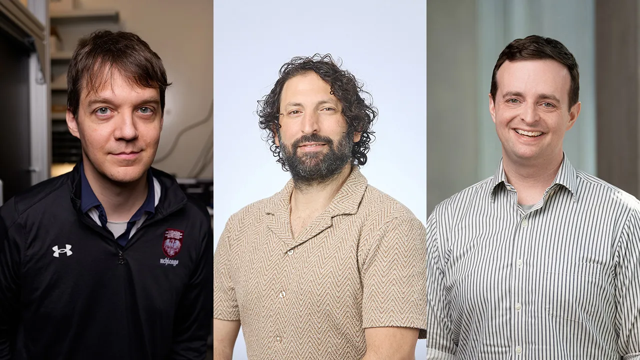
UChicago Pritzker School of Molecular Engineering researchers (from left) Assoc. Prof. Peter Maurer, Uri Zvi, PhD'25, and Prof. Aaron Esser-Kahn are co-authors of a paper that overturns a major assumption limiting quantum biosensors' effectiveness. (Photos courtesy of Jason Smith, Uri Zvi)
Inserting a diamond-based quantum sensor into a living cell can obtain previously unseen levels of detail about how life works and how diseases form.
“Think of it as an EKG for a single cell — a way to capture everything happening inside at once, in real time,” said University of Chicago Pritzker School of Molecular Engineering Prof. Aaron Esser-Kahn. “We routinely monitor vital signs in people — heart rate, breathing, temperature — but until now, there simply hasn’t been an equivalent way to take a cell’s vitals.”
An interdisciplinary research team from UChicago PME and the University of Iowa, originally aimed at making diamond-based quantum biosensors less toxic, less inflammatory, and overall easier on cells, unexpectedly revealed new insights that overturned a major assumption limiting the sensors' effectiveness.
“We found a missing piece of the puzzle,” said Uri Zvi, PhD’25, first author of the paper published in Advanced Materials.
When inserted into living cells, diamond-based biosensors experience energy level shifts called “zero-field splitting” (ZFS), which reduce the sensors’ effectiveness. It had always been assumed that these shifts were the diamond reacting to the cell’s temperature.
“ZFS of qubits in diamond is known to be impacted by temperature. So when people observed ZFS shifts they assumed that these were caused by local temperature changes, i.e., caused by cellular activities,” said UChicago PME Assoc. Prof. Peter Maurer, co-corresponding author of the new work along with Esser-Kahn and University of Iowa Asst. Prof. Denis Candido.
Instead, they discovered the diamond’s surface was causing the shifts. This greater understanding can be harnessed to get even more detailed readings of cells, Maruer said.
“This paper makes two major advancements,” Maurer said. “First, it resolves a longstanding discussion around the origin of observed shifts in qubit resonances in diamond nanoparticles, which have previously been associated with thermogenesis caused by cellular activity. We showed that these shifts are caused not by temperature but by a surface effect. Second, we showed that the observed qubit resonance shifts can be connected to cellular processes and could be an interesting proxy for probing cellular activity."
Following the clues
A previous interdisciplinary breakthrough both led to this mystery and gave the team the tools to crack it.
Last year, Zvi, immunoengineer Esser-Kahn, quantum engineer Maurer and theoretical physicist Candido collaborated on a new shell-encased biosensor inspired by the technology used in quantum dot LED television sets.
They found that coating a sensor in silica, intended only to make the diamond easier on the cell, also created a vastly more stable sensor. Their subsequent investigation found two things that Zvi said led directly to this new work.
First, the coating was dispersing electrons from the diamond’s center, inadvertently closing traps that had been causing spin noise. That was the reason for the increased performance, Zvi said. Second, this electron loss was flipping the charge of just the inside of the diamond from negative to positive.
“The moment we saw that, we started thinking, ‘Hey, this is a stark change,’” Zvi said.
This opened up the possibility that electric field effects were more impactful than thought, causing them to revise their theory. But the coated sensor also gave them the ability to test this experimentally. They could control for surface effects.
The cell’s temperature would be the same whether the sensor was coated or not, so if temperature were causing the ZFS shifts, both sensors would see them. Instead, they found the silica coating was suppressing the ZFS shifts. The surface was to blame.
“Then came the big piece of information,” Zvi said. “We were measuring a 5-degree change over 30 minutes inside of the cell. It's just not thermodynamically feasible.”
‘Unexplained and puzzling’
“I am far from an expert in biology, but a sudden cellular temperature variation of 4 to 5 degrees Celsius is very unexpected," Candido joked. “As a theorist, I always love it when experimentalists have unexplained and puzzling data, so it was very exciting!”
While the current research indicates that temperature was not responsible for the observed zero-field splitting shifts, that conclusion stands in stark contrast to much of the previous literature. Earlier studies had interpreted similar zero-field splitting shifts as evidence of intracellular temperature changes of 1 to 10 degrees, Zvi said.
This caused Zvi and Candido to go back to the theoretical drawing board. Not only did they find the true culprit behind the ZFS shifts, they also found an important way to distinguish between the variations of temperature and the electric field signal.
Future researchers can harness this to make more accurate temperature readings of cells and better biosensors to understand life itself.
“You can think about being able to follow whether a T cell is becoming a regulatory T cell or a killer T cell, whether a cell becomes a cancer cell or just a healthy cell,” Zvi said. “There is an entire new world to explore, and entirely new ways to explore it.”
Citation: "Probing Cellular Activity Via Charge-Sensitive Quantum Nanoprobes," Zvi et al, Advanced Materials, February 4, 2026. DOI: 10.1002/adma.202505107
Related News
-
New open-source software predicts energy transfer between tiny defects in solid materials
July 30, 2026The performance of many next-generation devices depends on controlling how energy flows at extremely small scales. In the field of microelectronics — where devices continue to shrink and new materials... -

Unlocking new quantum architectures
July 30, 2026A less-explored form of quantum code could be more powerful – and more stable – than its popular alternative in error correction. -

Engineering the Summer: Building tomorrow’s quantum computers
July 22, 2026Engineering the Summer is an annual series following UChicago Pritzker School of Molecular Engineering students as they embark on summer internships and career experiences. University of Chicago...